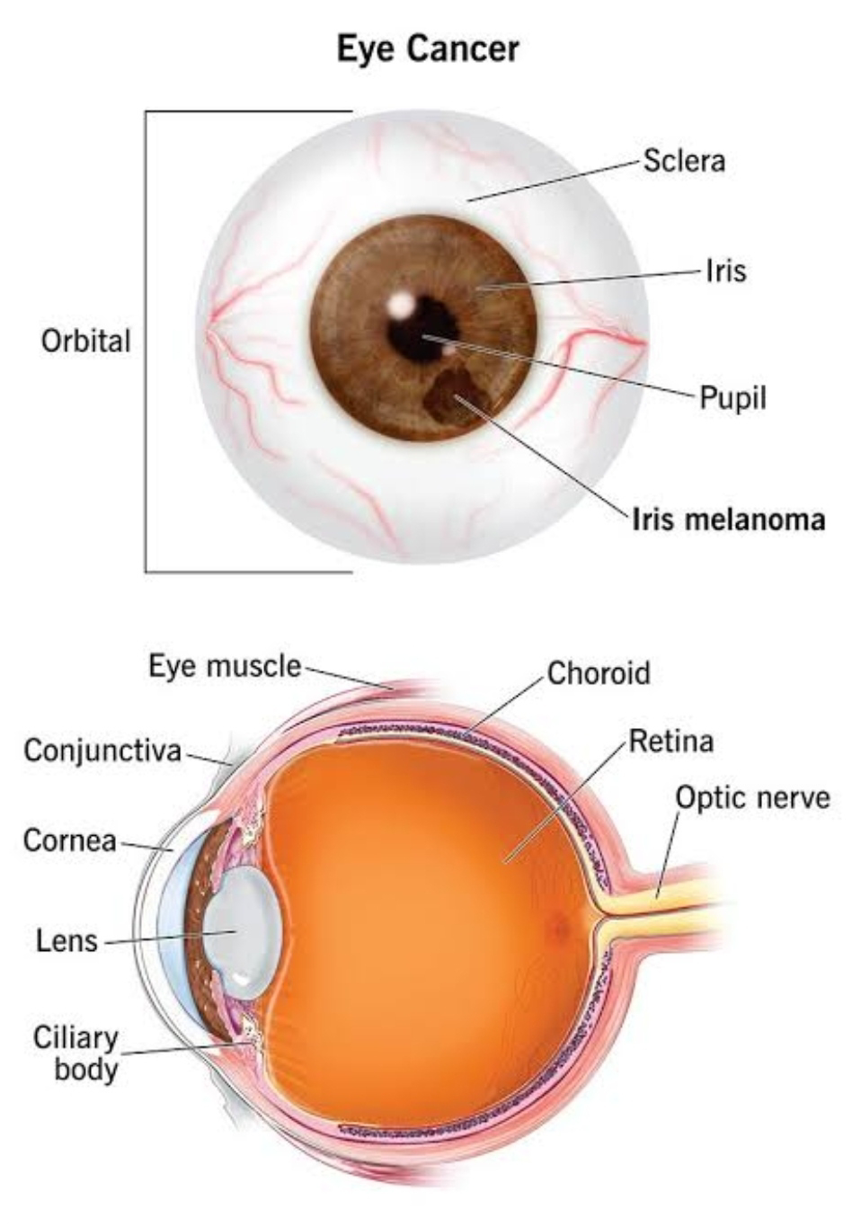
Eye tumor is otherwise known as ocular tumors. It is an abnormal grows in the eye, and it can be malignant or benign.
The most common type of eye tumor is metastatic, caused by cancer that has spread from one part of the body to another, often coming from the lung, breast, bowel, or prostate.
ETIOLOGY AND RISK FACTORS :-
1.over the age of 50 years, and rare in children
2.Common in White people and less common in Black people.
3.Men and women are equally affected by intraocular melanoma.
4.Medical history: Basal cell carcinoma, squamous carcinoma, sebaceous carcinoma, and malignant melanoma are all types of eyelid cancers. People who have extra pigmentation of the eye or skin around the eye, spots like moles in the eye, or multiple flat moles that are irregular in shape or color are more likely to develop intraocular melanoma.
5.Family history: Intraocular melanoma also sometimes runs in families.
6.Genetic mutation: Retinoblastoma is an eye cancer that affects young children and is caused by a genetic mutation.
7.Sunlight or certain chemicals may increase the risk of intraocular melanoma development.
TYPES OF EYE CANCER :-
1.Eye or intraocular melanoma-conjunctival, choroidal
2.Squamous cell carcinoma
3.Lymphoma or primary intraocular lymphoma-often associated with lymphoma in the brain.
4.Retinoblastoma-common in children, most common metastatic tumors, in the eye are from breast and lung cancers.
CLINICAL MANIFESTATIONS :-
1.Flashes of light, or floaters
2.Blurred vision, haloes, and shadows around images, especially of bright light.
3.Eye moles, such as skin moles, develop when certain cells grow together in a group, an 4.Abnormal brown spot on or in eye
of the eye that increases in size get angry looking blood vessels around.
5.Decrease in vision may be associated with pain
6.Bulging of one eye, or both, called proptosis
7.A lump or tumor on eyelid or in eye that is increasing in size, getting blood vessels.
8.Change in color of the iris
9.White reflex in the pupil
10.Eye tumor may first appear as a dark spot on the iris, the colored part of eye.
DIAGNOSTIC EVALUATION :-
1.History collection
2.Physical examination helps to find out like extra pigmentation of the eye or skin around the eye, spots like moles in the eye, or multiple flat moles.
3.Eye examination-examine any enlarged blood vessels on the outside of the eye is usually a sign of a tumor inside eye, look deep inside the eye with the help of a binocular indirect ophthalmoscope.
4.Slit-lamp-used to view the interior structures of the eye.
5.Eye ultrasound-used to produce images of the inside of the eye.
6.Optical coherence tomography-used to create pictures of the inside of the eye.
7.Fluorescein angiography-fluorescein is injected into the vein. The dye moves through body and into the blood vessels in the back of the eye, allowing to take pictures and view the inside the eye deeply.
MANAGEMENT :-
Aim of the treatment to reduce the risk of spreading, preserving the functional and structural integrity of the eye, preserving the cosmetic appearance of the eyeball and surrounding areas and to maintain the health and vision of the eye.
1.Limited resection - removal of the cancer affected part of the eye, e.g., iridectomy, choroidectomy, eyelid resection etc.
2.Enucleation - removal of the eyeball, in which the eyelids and eye muscles are not removed. Later, fit an artificial eye
3.Evisceration - partial removal of the eye contents, leaving the sclera or the white part of the eye behind.
4.Exenteration - removal of the eye, all orbital contents, and a specially designed prosthesis can later be fitted to preserve the appearance of the face.
5.Chemotherapy - involves the use of special drugs which may be administered orally or by injection to fight the cancer cells.
6.Radiation therapy is used to destroy cancer cells.
7.Plaque therapy or brachytherapy-a plate impregnated with the therapeutic agent is used to direct the treatment to the specific location alone, minimizing damage to surrounding tissue.
8.Laser therapy-uses lasers to shrink tumors.


0 Comments